22
3 B S c i e n t i f i c ® M i c r o s c o p y
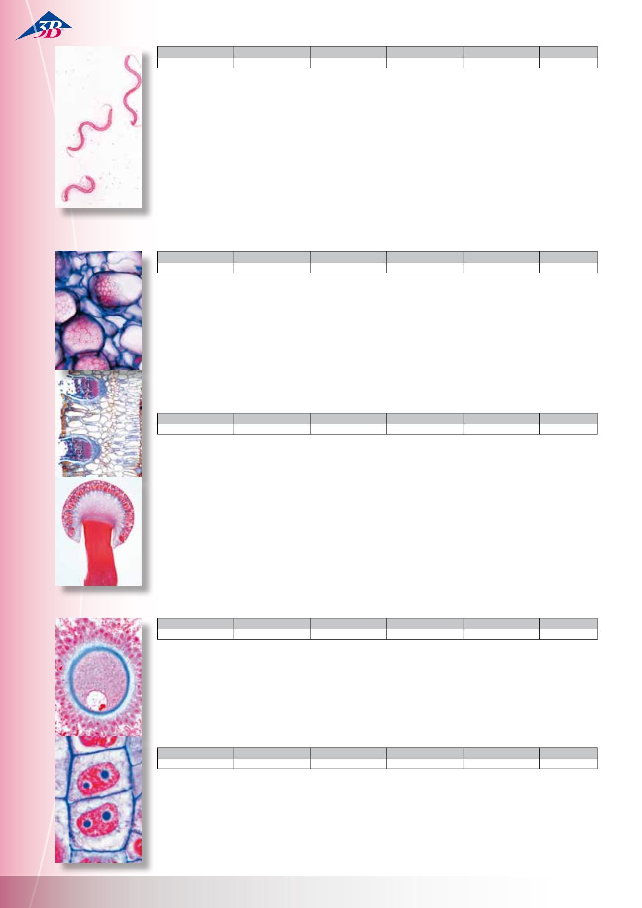
Bacteria Basis Set
25 Microscope Slides
The most important pathogenic and non-pathsgenic bacteria
1(d). Staphylococcus aureus, pus organism 2(d). Sarcina lutea,
chromogenic rods 3(e). Streptococcus pyogenes, pus organism
4(d). Streptococcus lactis, milk souring organism 5(d). Bacillus
subtilis, hay bacillus, smear with bacilli and spores 6(d). Bacillus
mycoides, soil organis 7(e). Bacillus anthracis, wool sorters
disease 8(e). Mycobacterium tuberculosis, tuberculosis 9(d).
Corynebacterium diphtheriae, diphtheria 10(e). Bacterium erysi-
pelatos, red murrain 11(d). Rhizobium radicicola, nitrogen fixing
bacteria 12(d). Proteus vulgaris, putrefaction 13(d). Escherichia
coli, colon bacteria 14(d). Eberthella typhi, typhoid fever 15(d).
Salmonella paratyphi, paratyphoid fever 16(f). Vibrio comma,
Asiatic cholera 17(d). Shigella dysenteriae, bacillary dysentery
18(d). Hemophilus influenzae, Pfeiffer bacillus 19(e). Spirillum
volutans, from putrid water 20(d). Rhodospirillum rubrum,
chromogenic spirilli 21(e). Clostridium botulinum (botulism),
food poisoning 22(g). Spirochaeta duttoni (Borrelia recurrentis),
in blood smear 23(d). Bacteria from mouth, with Gram positive
and negative rods 24(d). Bacteria from bread 25(d). Bacteria from
cheese.
W13011
W13040
W13011F
W13011S
W13011P
German
English
French
Spanish
Portuguese
BOTANY
Phanerogamae, Elementary Set
25 Microscope Slides
1(c). Simple plant cells, epidermis of Allium w.m. 2(d). Cell divi-
sion (mitosis) all stages, in Allium root tips l.s. 3(c). Starch grains,
t.s. of potato tuber 4(c). Cork cells, t.s. of bark of Quercus 5(d).
Stone cells, t.s. of fruit of pear 6(d). Root hairs on root tip 7(c).
Zea mays, corn, typical monocot root t.s. 8(c). Ranunculus, but-
tercup, typical dicot root t.s. 9(c). Zea mays, corn, monocot stem
t.s. 10(c). Triticum, wheat, gramineous stem t.s. 11(c). Aristolo-
chia, birthwort, one year stem t.s. 12(c). Aristolochia, older stem
t.s. 13(d). Cucurbita, pumpkin, stem with bundles and sieve tubes
l.s. 14(c). Sambucus, elderberry, stem with lenticels t.s. 15(c).
Tulipa, tulip, leaf epidermis with stomata w.m. 16(c). Zea mays,
corn, leaf t.s., monocot gramineous leaf 17(c). Syringa, lilac, leaf
t.s., dicot leaf 18(c). Fagus, beech, leaf bud t.s. shows leaf origin
19(d). Lilium, lily, flower bud t.s. shows flower diagram 20(d). Lili-
um, anthers t.s. shows pollen chambers and pollen grains 21(d).
Lilium, ovary t.s. with embryosac 22(e). Lilium, stigma with pol-
len and pollen tubes l.s. 23(c). Pinus, pine, leaf (needle) t.s. 24(d).
Triticum, wheat, grain (semen) t.s. with embryo and endosperm
25(d). Capsella, shepherd’s purse, l.s. of embryos in situ.
Fungi and Lichen
20 Microscope Slides
Phycomycetes 1(c). Mucor mucedo, w.m. of hyphae showing
sporangia 2(d). Rhizopus nigricans, w.m. of hyphae with deve-
loping zygotes (d). Synchytrium endobioticum, potato black
wart, t.s. of infected tissue 4(c). Plasmodiophora, t.s. of cabbage
rot Ascomycetes 5(c). Claviceps purpurea, t.s. of sclerotium 6(c).
Tuber rufum, truffle, t.s. of fruiting body showing asci 7(c). Peziza
sp., cup-fungus, t.s. of fruiting body with asci 8(d). Erysiphe
sp., mildew, t.s. of leaf with perithecia 9(d). Penicillium sp.,
blue mould on orange-rind, t.s. of hyphae with conidiophores
10(c). Aspergillus glaucum, brown-mould, w.m. of hyphae with
sporangia 11(b). Saccharomyces sp., yeast, budding, w.m. 12(d).
Taphrina pruni (Exoascus pruni), plum pockets, t.s. with haustoria
and asci Basidiomycetes 13(d). Puccinia graminis, t.s. of uredinia
on wheat 14(d). Puccinia graminis, wheat rust, t.s. of aecidia on
infected barberry leaf 15(d). Ustilago zeae, corn smut, infected
tissue, t.s. 16(c). Psalliota sp., mushroom, l.s. through pileus and
lamellae 17(c). Boletus edulis, pore fungus, l.s. through pores
18(c). Lycoperdon gemmatum, puff-ball, t.s. of fruiting body
Lichens 19(d). Xanthoria, lichen, t.s. of thallus showing hyphae
with symbiotic algae 20(d). Xanthoria, t.s. of apothecium.
The Animal Cell
12 Microscope Slides
1(c). Squamous epithelium, isolated cells from human mouth
2(d). Striated muscle l.s. showing nuclei, striations 3(d). Compact
bone and hyaline cartilage t.s., two sections for comparison 4(e).
Nerve fibres isolated, fixed and stained by osmic acid to show
myelin sheaths and Ranvier’s nodes 5(d). Liver of Salamandra
t.s., simple animal cells 6(f). Kidney of mouse, t.s. vital stained
to demonstrate storage 7(d). Ovary of cat, t.s. showing primary,
secondary, and Graafian follicles 8(d). Testis of frog, t.s. showing
spermatogenesis 9(e). Salamandra larva, t.s. of skin and other or-
gans selected to show cell division (mitosis) 10(f). Uteri of Ascaris
megalocephala, t.s. stained to show meiosis with chromosomes
and nuclear spindles 11(f). Salivary gland of Chironomus larva.
Giant chromosomes showing large chromomeres. Stained for
DNA after Feulgen 12(e). Ova from Psammechinus (sea urchin).
Unfertilized ova, fertilized ova, early cleavage stages.
CYTOLOGY AND EMBRYOLOGY
Plant Cell
12 Microscope Slides
1(c). Epidermis of Allium (onion), w.m. showing simple plant cells
with cell walls, nuclei and cytoplasm 2(d). Root tips of Allium
cepa l.s. showing cell division (mitosis) in all stages 3(e). Pollen
mother cells of Lilium. Prophase of first maturation division
(meiosis) 4(f). Pollen mother cells of Lilium. Metaphase and ana-
phase of first maturation division 5(c). Wood of Tilia macerated
and w.m. 6(d). Fruit of Pyrus (pear) t.s. showing stone cells 7(c).
Tuber of Solanum (potato) t.s. shows cork and starch grains 8(d).
Cucurbita pepo (pumpkin) l.s. of stem showing vascular bundles
with sieve tubes, spiral and annular vessels 9(c). Ricinus endo-
sperm t.s. showing aleurone grains 10(d). Anthers of Lilium (lily),
t.s. pollen sacs and pollen grains 11(d). Ovary of Lilium (lily), t.s.
arrangement of ovules and embryosac 12(e). Spirogyra showing
conjugation stages and zygotes.
W13328
W13428
W13328F
W13011S
W13328P
German
English
French
Spanish
Portuguese
W13013
W13042
W13013F
W13013S
W13013P
German
English
French
Spanish
Portuguese
W13023
W13052
W13023F
W13023S
W13023P
German
English
French
Spanish
Portuguese
W13024
W13053
W13024F
W13024S
W13024P
German
English
French
Spanish
Portuguese
M i c r o s c o p e S l i d e s


