K13
K24
K23
F16
F16
K24
K23
K13
. . . g o i n g o n e s t e p f u r t h e r
31
3B MICRO
anatomy
™ Kidney
This extremely detailed model shows the morphologic/functional units of
the kidney greatly magnified. Six model zones illustrate the following fine-
tissue structures that serve the production of urine:
• Longitudinal section of a kidney
• Section of renal cortex and renal medulla
• Wedge-shaped section of a kidney lobe with a diagrammatic depiction of
three nephrons with Henle’s loops of different lengths and diagrammatic
depiction of the vascular supply
• Diagrammatic illustration of a nephron with a short Henle’s loop and
didactic/diagrammatic illustration of the vascular supply
• Diagrammatic illustration of an opened renal corpuscle with nephron
and light-microscopic transverse sections of the proximal, attenuated and
distal segments of a renal tubule
• Diagrammatic/didactic illustration of an opened renal corpuscle
Mounted on a base.
23.5x25.5x19 cm; 1.3 kg
L/E/D/S/F/P/I/J www.
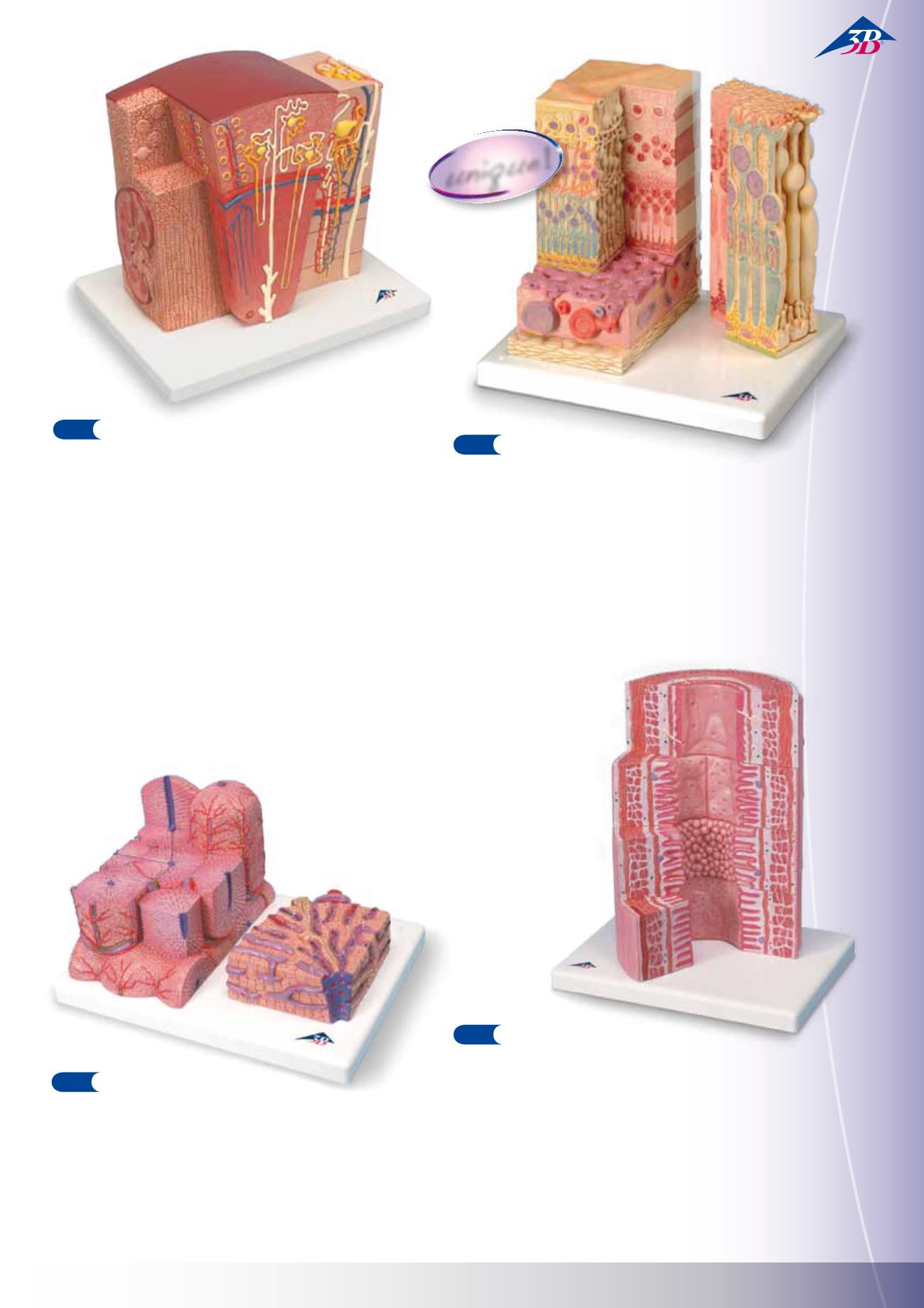
3B MICRO
anatomy
™ Digestive System
The model illustrates the structure of the fine tissues of four characteristic
sections of the digestive system: oesophagus, stomach, small intestine,
large intestine.
The front of the model, from top to bottom, shows a magnified view in his-
tological section of the individual sections of the digestive system and their
fine tissue structures.On the back of the model, highly magnified views of
didactically interesting areas of each of the digestive system sections shown
on the front are emphasized.
29.5x26x18.5 cm; 1.5 kg
L/E/D/S/F/P/I/J www.
3B MICRO
anatomy
™ Liver
This 2-part model shows a highly magnified diagrammatic view of a section
of the liver. The left part of the model shows a section of the liver that
comprises several lobules. The right part of the model is a highly magnified
view of the sectioned lobule on the left.
15x26x18.5 cm; 0.7 kg
L/E/D/S/F/P/I/J www.
3B MICRO
anatomy
™ Eye
This model illustrates the microscopic structure of the retina with choroid
and sclera. The left block-like, layered side of the model side shows the
complete structure of the retina including the vascular layer and parts of
the sclera from a light microscopic view. The right part of the model is a
sectional enlargement. It shows the microscopic structure of the photore-
ceptors and the cells of the pigmented layer.
25x23x18.5 cm; 1.2 kg
L/D/E/F/S/P/I/J www.
unique
!
3 B M I C RO
a n a t omy
™


