. . . g o i n g o n e s t e p f u r t h e r
23
Set of Genetic Slides
25 Microscope Slides
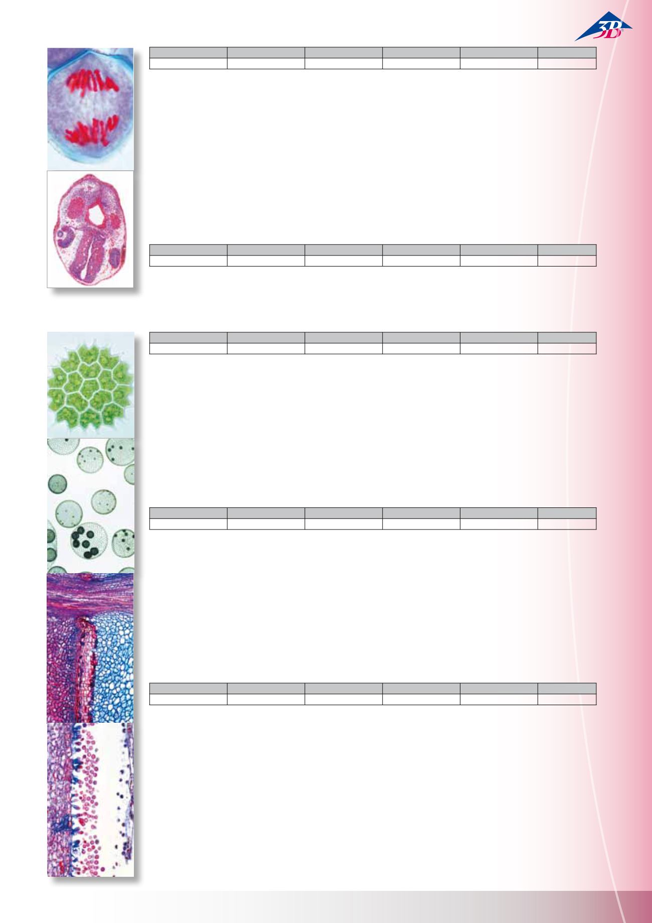
1(d). Allium, root tips, l.s. showing all stages of mitosis 2(e). Esch-
scholtzia, stigma, w.m. showing penetrating pollen 3(e). Lilium,
microspore mother cells, first division, leptotene to zygotene
4(e). Lilium, first division, diakinesis to telophase 5(f). Lilium,
second division, interkinesis to tetrad stage 6(f). Polytrichum,
moss, archegonium, w.m. 7(f). Polytrichum, moss, archegoni-
um, l.s. 8(e). Spirogyra scalariform conjugation showing zygotes
following conjugation 9(d). Sea urchin, developing of eggs, w.m.
of most stages up to pluteus 10(f). Giant chromosomes from
salivary gland of Chironomus, squash preparation stained for
chromomeres 11(f). Giant chromosomes, section 12(e). Ascaris,
fertilisation of eggs, t.s. 13(f). Ascaris, male and female pronuclei,
t.s. 14(f). Ascaris, meiosis and early cleavage, t.s. 15(e). Testis of
crayfish, t.s. showing meiosis 16(d). Testis of mouse, t.s. showing
spermatogenesis 17(d). Ovary of rabbit, l.s. showing follicles in
various stages 18(f). Embryology of fish, l.s. of embryo showing
animal mitosis 19(h). Chromosomes, human, female, of culture
of peripheral blood 20(i). Chromosomes, human, male, of culture
of peripheral blood 21(f). Drosophila genetics, adult wild type,
w.m. 22(f). Drosophila genetics, “barr eye” mutant, w.m. 23(f).
Drosophila genetics, “brown eye” mutant, w.m. 24(f). Drosophila
genetics, “vestigial wing” mutant, w.m. 25(f). Drosophila genetics,
“white eye” mutant, w.m.
Frog Embryology (Rana)
10 preparations with accompanying guide. For details, please go to
ECOLOGY AND ENVIRONMENT
The Microscopic Life in the Water
25 Microscope Slides
1(e). Amoeba proteus, amoeba 2(c). Ceratium hirundinella, di-
noflagellates 3(c). Euglena, green flagellate with eyespot 4(d). Ra-
diolaria, marine rhizopods 5(c). Paramecium, nuclei stained 6(d).
Stylonychia, a common ciliate 7(b). Spongilla, fresh water sponge,
isolated spicules 8(d). Hydra, w.m. or section 9(d). Rotatoria, roti-
fers, mixed species 10(c). Daphnia, water flea, a phyllopod 11(c).
Cyclops, a copepod 12(d). Chironomus, gnat, larva w.m. 13(d).
Putrefaction causing bacteria from hay infusions 14(c). Oscilla-
toria, a filamentous blue green alga 15(c). Diatomeae, diatoms,
mixed species 16(d). Desmidiaceae, desmids, mixed species 17(c).
Spirogyra, green alga with spiral chloroplasts 18(d). Eudorina,
small colonies within gelatinous sheaths 19(c). Cladophora, green
alga, branched filaments 20(c). Draparnaldia, main filaments and
branchings 21(c). Microcystis, irregular colonies 22(c). Ulothrix,
green alga with girdle-shaped chloroplasts 23(d). Oedogonium,
vegetative filaments 24(e). Volvox, with daughter colonies and
sexual stages 25(d). Mesothaenium, rod-shaped desmids
The Forest, Consequences of Pollution
20 Microscope Slides
1(c). Pine (Pinus), healthy leaves, t.s. 2(c). Pine (Pinus) leaves
damaged by acid rain, t.s. 3(c). Fir (Abies), healthy leaves, t.s.
4(c). Fir (Abies), stem tip damaged t.s. 5(c). Beech (Fagus), healthy
leaves t.s. 6(c). Beech (Fagus), t.s. of leaves with destroyed
epidermis and chloroplasts 7(d). Rhytisma acerinum, tar spot of
maples, consequence of single-crop farming 8(d). Early leaf fall,
caused by thawing salt 9(d). Healthy lichen, indicator of clean
air 10(d). Damaged lichen, caused by air pollution 11(c). Healthy
wood of beech, t.s. 12(d). Wood destroyed by fungus 13(d).
Polyporus, wood rot fungus, fruiting body t.s. 14(d). Root nodules
of Alnus, with symbiotic bacteria 15(d). Spruce beetle (Cryphalus
picea), larva t.s. 16(c). Wood with normal annual rings, t.s. 17(c).
Wood with anomalous narrow annual rings caused by drought,
t.s. 18(d). Bark with larval galleries of spruce beetle, t.s. 19(d).
Pineapple-like gall on spruce caused by lice, t.s. 20(d). Gall nut on
oak caused by insects, t.s.
Water Pollution, Problems and Results
20 Microscope Slides
1(d). Intestinal bacteria (Escherichia coli) from putrid water 2(e).
Putrefactive bacteria (Spirillum) from sludge poor in oxygen 3(d).
Putrefactive bacteria (Sphaerotilus) bacteria, forming long chains
4(d). Sludge bacteria (Methanobacterium) causing sewer gas 5(d).
Sulphur bacteria (Thiocystis) 6(c). Wasserbluthe (Microcystis),
blue-green alga “blooming” in stagnant water 7(c). Anabaena,
blue green algae, in eutrophic water 8(c). Spirogyra, filamentous
green alga in nutrient-rich water 9(d). Spirulina, corkscrew-
shaped algae occurring in bitter seas 10(c). Chlamydomonas,
one-celled green alga in eutrophic water 11(c). Cladophora, green
alga from moderately polluted water 12(c). Diatoms, mixed algae
from scarcely polluted water 13(c). Euglena, green flagellates
occurring in stagnant eutrophic water 14(d). Ciliates, different
species from nutrient-rich water 15(d). Rotifers (Rotatoria), small
animals from putrid water 16(d). Tubifex, fresh water oligochae-
te, living in the sludge 17(d). Carchesium, stalked ciliate from mo-
derately polluted water 18(d). Water mold (Saprolegnia), harmful
to plants and animals 19(d). Skin of fish injured by chemicals, t.s.
20(d). Skin ulcer of an amphibian, t.s.
W13025
W13054
W13025F
W13025S
W13025P
German
English
French
Spanish
Portuguese
W13335
W13435
W13335F
W13335S
W13335P
German
English
French
Spanish
Portuguese
W13331
W13431
W13331F
W13331S
W13331P
German
English
French
Spanish
Portuguese
W13332
W13432
W13332F
W13332S
W13332P
German
English
French
Spanish
Portuguese
W13027
W13056
W13027F
W13027S
W13027P
German
English
French
Spanish
Portuguese
M i c r o s c o p e S l i d e s


