B60
D17
G42
A79
A79
G42
D17
B60
30
3 B S c i e n t i f i c ® M i c r o s c o p y
3B MICRO
anatomy
™ Muscle Fibre
The model illustrates a section of a skeletal muscle fibre and its neuromus-
cular end plate magnified approx. 10,000 times. The muscle fibre is the
basic element of the diagonally striped skeletal muscle.
23.5x26x18.5 cm; 1.1 kg
L/E/D/S/F/P/I/J
3B MICRO
anatomy
™ Tongue
The latest model in our 3B MICRO
anatomy
™ series, the tongue, is fasci-
nating in that it combines various enlargements of specific parts of the
tongue in one model. It comprises a macroscopic view of the tongue in
life size (dorsal view) and microscopic views of the various papillae of the
tongue (10-20x life size) and of a taste bud (approx. 450x life size). All views
are mounted on a base that also features an overview of the sensory and
sensitive innervation of the tongue. A unique model for an intensive study
of the tongue.
14,5x32,5x20 cm; 0.8 kg
L/D/E/F/I/S/P/J/R/C
3B MICRO
anatomy
™ Artery and Vein
The model shows a medium-sized muscular artery with two adjacent veins
from the antebrachial area with adjoining fat tissue and muscle enlarged
14 times. The model illustrates the reciprocal anatomical relationship of
artery and vein and the basic functional techniques of the venous valves
(“valve function” and “muscle pump”). The left vein and the middle artery
are fenestrated in the upper anterior segment, revealing the various layers
of the wall structure in a cross and longitudinal section and in top view.
The right vein is opened throughout in the anterior segment, revealing the
orifice of a feeder vein and two venous valves, i.e. “flap valves” formed by
a duplication of the tunica intima. On the rear of the model, the relief of
two veins is shown to illustrate the functional aspect of the venous valves.
Supplied on base.
26x19x18.5 cm; 0.9 kg
L/D/E/S/F/P/I/J
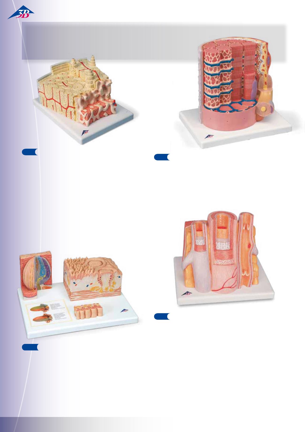
3B MICRO
anatomy
™ Bone Structure
This extremely detailed model depicts a three dimensional section of a
lamellar bone, showing the typical structure of a tubular bone enlarged
80 times. Various planes are shown in cross and longitudinal section
through all levels of the bone, as well as a 2‑planesection through the
inner structure of the bone marrow. The typical elements of a lamel-
lar bone are easily identified and help to understand its structure and
function with the characteristic osteons, also referred to as Haversian
systems. This model allows a graphic illustration of the interplay of the
individual components, such as spongy and compact substance, endos-
teum, cortical substance, osteocytes, Volkmann and Haversian canals.
Supplied on base.
26x19x14.5 cm; 0.8 kg
E/D/S/F/P/J www.
3 B M I C RO™
FURTHER ITEMS FOR YOUR BIOLOGY LESSON…
As world leader in anatomical model production, we would also
like to show you our most popular models.


