20
3 B S c i e n t i f i c ® M i c r o s c o p y
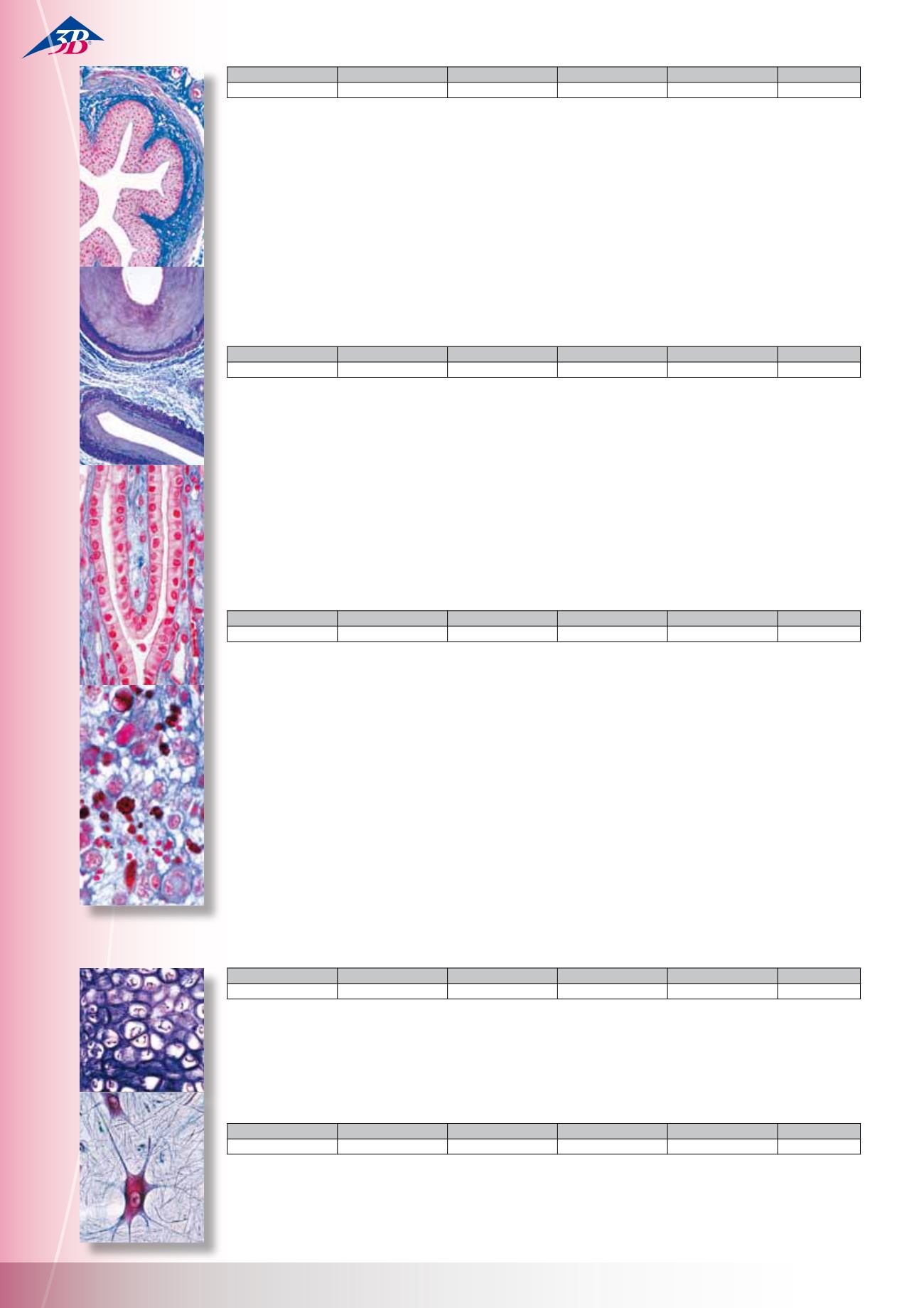
Normal Human Histology, Basic Set
40 Microscope Slides
When compiling the series only top quality, histologically
fixed material was used for the preparation of the slides. The
cutting thickness of the microtome sections is normally 6 – 8
mm. The use of special staining methods guarantees a clear,
multicoloured representation of all tissue structures. This slide
series occupies a special position due both to the quality of the
original material and the case taken during it‘s preparation.
1(c). Squamous epithelium, human, isolated cells 2(f). Areolar
connective tissue, human w.m. 3(f). Hyaline cartilage, human
t.s. 4(f). Compact bone, human t.s. 5(f). Striated muscle, human
l.s. 6(f). Heart muscle, human l.s. and t.s. 7(f). Artery, human t.s.
8(f). Vein, human t.s. 9(f). Lung, human t.s. 10(c). Blood smear,
human 11(f). Spleen, human t.s. 12(f). Thyroid gland, human t.s.
13(f). Thymus gland from human child t.s. 14(f). Tongue, human
t.s. 15(f). Tooth, human l.s. 16(f). Parotid, human gland t.s. 17(f).
Oesophagus, human t.s. 18(f). Stomach, human, fundic region
t.s. 19(f). Duodenum, human t.s. (small intestine) 20(f). Colon,
human t.s. (large intestine) 21(f). Pancreas, human t.s. 22(f).
Liver, human t.s. 23(e). Vermiform appendix, human t.s. 24(f).
Kidney, human t.s. 25(f). Adrenal (suprarenal) gland, human t.s.
26(f). Ovary, human t.s. 27(f). Uterus, human t.s. 28(f). Placenta,
human t.s. 29(f). Testis, human t.s. 30(f). Epididymis, human t.s.
31(f). Cerebrum, human t.s. 32(f). Cerebellum, human t.s. 33(f).
Spinal cord, human t.s34(f). Sympathetic ganglion, human t.s.
35(e). Skin of palm, human t.s. 36(e). Scalp, human, l.s. of hair
follicles 37(e). Scalp, human, t.s. of hair follicles 38(f). Retina, hu-
man t.s. 39(e). Finger tip from foetus with nail development l.s.
Normal Human Histology, Large Set, Part I.
50 Microscope Slides
1(c). Isolated squamous epithelium, human 2(e). Connective tissue,
human, sec. 3(e). Columnar epithelium, human gall bladder, t.s.
4(e). Ciliated epithelium, human trachea, t.s. 5(e). Smooth muscles,
human, l.s. and t.s. 6(e). Striated muscles, human, l.s. 7(e). Heart
muscles, human, l.s. and t.s. 8(e). Hyaline cartilage, human, sec.
9(e). Elastic cartilage of epiglottis, human, t.s. 10(e). Bone, compact
substance, human, t.s. 11(e). White fibrous tissue (tendon), human,
l.s. 12(e). Red bone marrow, human, t.s. 13(d). Scalp, human, l.s.
of hair follicles 14(e). Artery, human, t.s. 15(e). Vein, human, t.s.
16(c). Blood smear, human, Giemsa stain 17(e). Lung, human, t.s.
18(f). Larynx of human foetus, t.s. 19(e). Lymph gland, human, t.s.
20(e). Thyroid gland, human, t.s. 21(f). Pituitary gland, human,
t.s. 22(e). Spleen, human, t.s. 23(e). Tongue, human, t.s. 24(e).
Oesophagus, human, t.s. 25(e). Sublingual gland, human, t.s. 26(e).
Stomach, pyloric region, human, t.s. 27(e). Pancreas, human, t.s.
28(e). Small intestine, human, t.s. 29(e). Large intestine, human,
t.s. 30(e). Liver, human, t.s. 31(e). Kidney, human, t.s. 32(f). Adrenal
gland, human, t.s. 33(e). Ureter, human, t.s. 34(e). Urinary bladder,
human, t.s. 35(f). Ovary, human, t.s. 36(e). Uterus, human, t.s.
37(e). Uterine tube, human, t.s. 38(e). Placenta, human, t.s. 39(e).
Umbilical cord, human, t.s. 40(e). Mammary gland, human, sec.
41(f). Testis, human, t.s. 42(e). Epididymis, human, t.s. 43(f).
Olfactory epithelium, human, t.s. 44(f). Retina, human, t.s. 45(g).
Internal ear, human foetal, t.s. 46(f). Touch corpuscles in human
skin, t.s. 47(e). Nerve, human, l.s. 48(e). Spinal cord, human, t.s.
49(e). Cerebellum, human, t.s. 50(e). Cerebrum, cortex, human, t.s.
Normal Human Histology, Large Set, Part II.
50 Microscope Slides
1(e). Soft palate, human t.s. 2(e). Adipose tissue, human, sec. stained for fat
3(f). White fibrous cartilage, human intervertebral disc, sec. 4(e). Striated
(skeletal) muscle, human t.s. 5(e). Spongy (cancellous) bone, human t.s.
6(e). Bone development, vertical l.s. of foetal skull-cap 7(e). Bone deve-
lopment, l.s. of foetal finger 8(e). Joint of human foetus, l.s. 9(e). Tooth,
human, t.s. of crown 10(f). Tooth, human, complete l.s. 11(f). Tooth deve-
lopment fromhuman foetus, l.s. 12(e). Aorta, human, t.s. routine stained
13(e). Trachea fromhuman foetus t.s. 14(f). Thymus fromhuman child, t.s.
15(f). Parathyroid gland (Gl. parathyreoidea), human t.s. 16(e). Tonsil (Ton-
silla palatina), human t.s. 17(e). Parotid gland (Gl. parotis), human t.s. 18(e).
Submaxillary gland (Gl. submandibularis), human t.s. 19(e). Stomach,
fundic region, human t.s. 20(e). Stomach, cardiac region, human t.s. 21(e).
Jejunum, human t.s. 22(f). Small intestine (Duodenum) t.s. colouring of
goblet cells, PAS-HE 23(e). Vermiformappendix, human t.s. 24(e). Rectum,
human t.s. 25(e). Gall bladder, human t.s. 26(e). Liver of human foetus sec.,
developing blood cells 27(e). Urethra, human, t.s. 28(e). Seminal vesicle
(Gl. vesiculosa), human t.s. 29(e). Spermatic cord (Ductus deferens), human
t.s. 30(e). Prostate, human, t.s. 31(e). Spermsmear, human 32(f). Corpus
luteum in t.s. of human ovary 33(e). Vagina, human t.s. 34(g). Cerebral cor-
tex, human, t.s. silvered (Golgi or Palmgren) 35(g). Cerebral cortex, human,
t.s. stained for neuroglial cells after Held 36(g). Cerebellum, human, t.s.
silvered (Golgi or Palmgren) 37(f). Thalamus, human, stained after KlŸver –
Barrera 38(f). Medulla oblongata, human, t.s. routine stained 39(g). Spinal
cord, human, t.s. silvered (Golgi or Palmgren) 40(f). Sympathetic ganglion,
human t.s. routine stained 41(e). Peripheral nerve, human t.s. 42(e). Optic
nerve, human t.s. 43(e). Cornea fromeye, human t.s. 44(e). Eyelid, human,
t.s. 45(e). Skin fromfinger tip, human, vertical l.s. 46(d). Scalp, human,
horizontal l.s. shows t.s. of hair follicles, 47(e). Nail development, sagittal
l.s. finger tip of human foetus 48(h). Human chromosomes in smear from
culture of blood, male 49(i). Human chromosomes in smear fromculture
of blood, female 50(f). Barr bodies (human sex chromatin) in smear from
female squamous epithelium
HI STOLOGY – COMPREHENS IVE SET
Tissues
15 Microscope Slides
1(c). Squamous epithelium, scrapings from human mouth, w.m.
2(e). Columnar epithelium, human gall bladder, t.s. 3(e). Ciliated
epithelium, human trachea, t.s. 4(d). Skin, human, from general
body surface showing sweat glands 5(d). Human scalp, l.s. of
hair 6(d). Developing of nail, human embryo, l.s. 7(e). Hyaline
cartilage, human, t.s. 8(d). Elastic cartilage, ear of pig, t.s. 9(e).
Developing cartilaginous bone, joint of human foetus, l.s. 10(e).
Compact bone, c.s. and l.s. 11(f). Striated muscle, human, l.s.,
staining of striations 12(e). Striated muscle, human, t.s. 13(e).
Smooth muscle, human, t.s. and l.s. 14(e). White fibrous tissue,
human tendon, l.s. 15(e). Adipose tissue, human, t.s.
Nervous System
11 Microscope Slides
1(e). Cerebrum, human, cortex, t.s. 2(e). Cerebellum, human,
t.s. 3(f). Cerebellum, human, t.s., Weigert stained 4(e). Spinal
cord, human, t.s. for general structure 5(e). Nerve, human, l.s.
6(e). Nerve, human, t.s. 7(f). Spinal cord, cat, t.s., KlŸver-Barrera
stained 8(e). Spinal cord, cow, t.s., Nissl stained 9(f). Cerebrum,
cat, t.s., Golgi stained 10(e). Brain, rat, median l.s. 11(d). Vertebra
with spinal cord, rat, t.s.
W13308
W13408
W13308F
W13308S
W13308P
German
English
French
Spanish
Portuguese
W13309
W13409
W13309F
W13309S
W13309P
German
English
French
Spanish
Portuguese
W13319
W13419
W133319F
W13319S
W13319P
German
English
French
Spanish
Portuguese
W13312
W13412
W13312F
W13312S
W13312P
German
English
French
Spanish
Portuguese
W13310
W13410
W13310F
W13310S
W13310P
German
English
French
Spanish
Portuguese
M i c r o s c o p e S l i d e s


