. . . g o i n g o n e s t e p f u r t h e r
19
SERIES FOR SECONDARY SCHOOLS
Series I. Cells, Tissues and Organs
13 Microscope Slides
1(d). Simple animal cells in sec. of salamander liver 2(d). Mitosis,
l.s. from Allium root tips 3(c). Ranunculus, buttercup, t.s. of a
typical dicot root 4(e). Monocot and dicot stems, two t.s. for com-
parison 5(c). Syringa, lilac, t.s. of a typical mesophytic dicot leaf
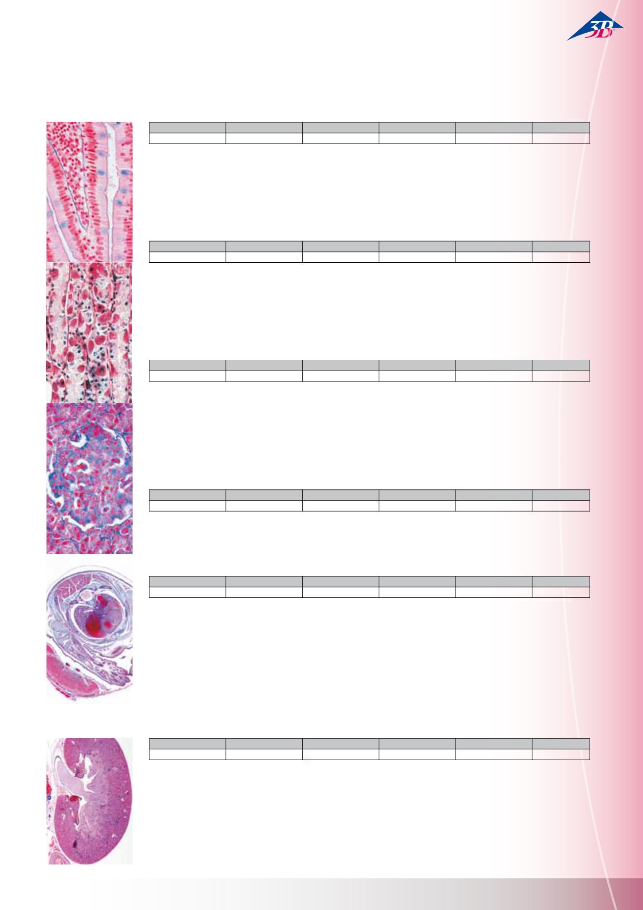
6(c). Columnar epithelium, t.s of blind gut from rabbit 7(e). Bone
and hyaline cartilage, t.s. 8(d). Striated muscles of mammal, l.s.
9(d). Smooth muscles of mammal, l.s. and t.s. 10(c). Lung of cat,
t.s. 11(c). Human blood smear 12(d). Human body skin, l.s. 13(f).
Young mouse, sag. s. of entire specimen for all structures.
Series II. Metabolism
15 Microscope Slides
1(e). Hydra, fresh water polyp, t.s. with ectoderm and entoderm
2(d). Carabus, ground beetle, gizzard 3(c). Salivary gland of cat,
t.s. 4(c). Oesophagus of cat, t.s. 5(d). Fundic stomach of cat, t.s.
6(c). Small intestine of cat, t.s. routine stained 7(f). Small intesti-
ne, t.s. blood vessels injected 8(d). Appendix of human, t.s. 9(c).
Large intestine of cat, t.s. 10(c). Liver of pig, t.s. 11(f). Malpighian
tubules of insect, t.s. 12(c). Primordial kidney (mesonephros) of
frog, t.s. 13(d). Hind-kidney (metanephros) of rabbit, t.s. 14(d).
Kidney of mouse with pelvis, l.s. 15(f). Kidney of mouse, t.s.
injected to show storage
Series III. Organs of Sense
16 Microscope Slides
1(e). Paramaecium, silvered to show the neuroformative system
2(d). Lumbricus, earthworm, t.s. with ventral nerve cord 3(e).
Insect brain, frontal l.s. 4(e). Planaria, sec. through ocelli 5(f).
Haliotis, marine snail, pinhole camera eye l.s. 6(e). Helix, snail,
eye l.s. 7(e). Alloteuthis, cuttlefish, camera eye l.s. 8(e). Com-
pound eye of an insect, l.s. 9(e). Young rat, head with eyes t.s.
10(d). Retina of cat, t.s. showing rods and cones 11(e). Internal
ear (cochlea) from guinea pig, l.s. 12(e). Taste buds from tongue
of rabbit, t.s. 13(e). Peripheral nerve fibres, osmic acid material
showing Ranvier’s nodes 14(c). Spinal cord of cat t.s. with large
motor nerve cells 15(c). Cerebellum of cat, t.s. routine stained
16(f). Cerebrum of cat, t.s. silvered to show the pyramid cells
Series IV. Hormone Organs and Hormonal Function
7 Microscope Slides
1(d). Ovary of cat, with follicles and corpus luteum t.s. 2(d). Testis
of mouse, t.s. showing Leydig’s cells 3(d). Adrenal (suprare-
nal) gland of cat, t.s. 4(d). Pancreas of cat, t.s. with islets of
Langerhans, 5(f). Thyroid gland, normal function t.s. 6(f). Thyroid
gland, over-activity of the gland t.s. 7(f). Hypophysis (pituitary
body) sagittal l.s.
Series V. Genetics, Reproduction and Embryology
19 Microscope Slides
1(g). DNA and RNA stained in different colours, l.s. onion root
tips 2(e). Lilium, young anthers, meiosis, early prophase stage,
t.s. 3(e). Lilium, young anthers, diplotene stage, t.s. 4(d). Lilium,
ovary with embryosac t.s. 5(d). Capsella bursa pastoris, l.s. of
embryos 6(h). Human chromosomes, spread in the metaphase
stage, w.m. 7(g). Lamp brush chromosomes 8(e). Hydra with testis
t.s. 9(e). Hydra with ovaries t.s. 10(f). Tapeworm (Taenia), mature
proglottid, w.m. 11(f). Ascaris, sec. of uteri showing maturation of
ova 12(e). Cockchafer (Melolontha), ovaries t.s. 13(d). Frog (Rana),
testis t.s. showing spermatogenesis 14(f). Frog embryology: four
cell stage t.s. 15(f). Frog: morula stage l.s. 16(f). Frog: neurula
stage t.s. 17(f). Chicken (Gallus) embryology: 24 hour t.s. 18(f).
Chicken embryology: 72 hour t.s. 19(d). Mouse, uterus containing
embryo t.s.
Histology of Mammalia, Elementary Set
25 Microscope Slides
1(c). Squamous epithelium, isolated cells 2(e). Fibrous connective
tissue, w.m. from pig mesentery 3(e). Adipose tissue of mammal,
fat stained 4(c). Hyaline cartilage of calf, t.s. 5(e). Compact bone
of cow, t.s. 6(d). Striated muscles of cat, l.s. 7(d). Smooth muscles
of cat, t.s. and l.s. 8(c). Blood smear, human 9(d). Artery of cat or
rabbit, t.s. 10(d). Vein of cat or rabbit, t.s. 11(c). Lung of cat, t.s.
12(c). Pancreas of pig with islets of Langerhans t.s. 13(c). Tongue
of cat, t.s. with cornified papillae 14(d). Stomach of cat, fundic
region t.s. 15(c). Small intestine of cat or rabbit, t.s. 16(d). Liver of
pig, t.s. 17(d). Kidney of cat, t.s. 18(d). Ovary of rabbit, t.s., deve-
loping follicles 19(d). Testis of mouse, t.s., spermatogenesis 20(d).
Cerebrum of cat, t.s. 21(d). Cerebellum of cat, t.s. 22(c). Spinal
cord of cat, t.s. 23(e). Nerve fibres isolated, Ranvier’s nodes 24(e).
Motor nerve cells, smear from spinal cord 25(d). Scalp, human,
l.s. of hair follicles
With the series for secondary school, you will receive microscope slide collections on the most popular areas. You can place important
topics “under the microscope”.
W13300
W13400
W13300F
W13300S
W13300P
German
English
French
Spanish
Portugese
W13301
W13401
W13301F
W13301S
W13301P
German
English
French
Spanish
Portugese
W13302
W13402
W13302F
W13302S
W13302P
German
English
French
Spanish
Portugese
W13303
W13403
W13303F
W13303S
W13303P
German
English
French
Spanish
Portugese
W13304
W13404
W13304F
W13304S
W13304P
German
English
French
Spanish
Portugese
W13306
W13406
W13306F
W13306S
W13306P
German
English
French
Spanish
Portugese
HI STOLOGY – Detai l Sets
M i c r o s c o p e S l i d e s
Ser i es for Secondar y School s


